Many viruses (HIV, measles, influenza, etc.) have a lipid envelope that protects their genetic information. The viral particle forms a lipid vesicle at the plasma membrane of the infected cell carrying the genetic information of the virus before it escapes by cleaving the newly formed viral membrane envelope from the host cell membrane, a process called membrane fission. Would it be possible to observe this phenomenon in order to observe membrane fission?
When a virus emerges from a cell, complex membrane remodeling, called budding is induced. The final step of budding includes the separation of the newly formed membrane-enveloped virus from the intact plasma membrane. This is catalyzed by spiral filaments made of proteins from the ESCRT machinery (Endosomal Sorting Complex Required for Transport ESCRT-III), which constrict the membrane to the point of fission with the help of the energy-providing ATPase VPS4B.
IRIG researchers have developed an
in vitro system to reconstitute ESCRT-III filaments. They then used both atomic force microscopy to monitor the evolution of the diameter of the spiral tubes in real time and electron microscopy to image filament constriction at higher resolution. This revealed that the details of VPS4B action at two steps: first, VPS4B reduces gradually the diameter of the filaments (constriction); secondly, VPS4B cleaves the filaments asymmetrically into two ends, with one end taking the form of a "dome-like" structure.
Thus, cleavage and dome formation have been suggested to constrain the membrane such that it ultimately leads to the release of the virus from the host cell.
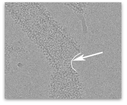
Structure of spiral tubes formed by ESCRT-III proteins.
The arrow shows where a tube is cut, with one of the two sides in the shape of a dome. Electron microscope image.
© CEA