Silver nanoparticles (Ag) are massively used for their antibacterial properties due to the release of silver in ionic form. Studies have estimated that we ingest between 1 and 80 μg of Ag per day. Moreover, medical devices such as catheters or burn dressings release silver nanoparticles into the bloodstream. To measure their impact on our health, researchers study the metabolism of silver nanoparticles in the liver. However, conventional human cell models do not allow for exhaustive
in vitro study of nanoparticles. On the other hand, animal models bring little insight into the cellular and molecular mechanisms involved.
Researchers at IRIG and ESRF Synchrotron in Grenoble, made use of an original 3D hepatocyte cellular model [1]. This bridges the gap between cellular and animal studies, by allowing for the observation
in vitro of phenomena occurring in vivo that standard 2D cellular models cannot reproduce, such as the excretion of transformed nanomaterials in the bile. By combining several characterization techniques, such as X-ray fluorescence nano-imaging, X-ray absorption spectroscopy and electron microscopy, the researchers studied the behavior of nanoparticles in the liver [2]. Hepatocytes, the major of liver cells, were exposed to silver nanoparticles on the one hand, and to a silver salt on the other, in order to simulate injection or ingestion, respectively. X-ray and 3D electron microscopy images revealed silver diffusion into various intracellular compartments (cytosol, nuclei, ...); the images also showed silver accumulation in vacuoles (Figure A white arrow), and excretion into bile-collecting ducts (Figure B white arrow). Speciation analyses revealed the formation of both organic silver-sulfur AgS species, as previously observed in 2D cell cultures [3] and the formation of inorganic Ag2S species not observed previously.
Using a pseudo-organ as cellular model and a customized combination of elemental and structural nano-imaging and atomic spectroscopy, this study reveals subcellular and molecular mechanisms linked to the human toxicity of a broadly used nanomaterial in the liver. These results allow us to better understand the effects on our health, and contribute to the development of safer nanomaterials.
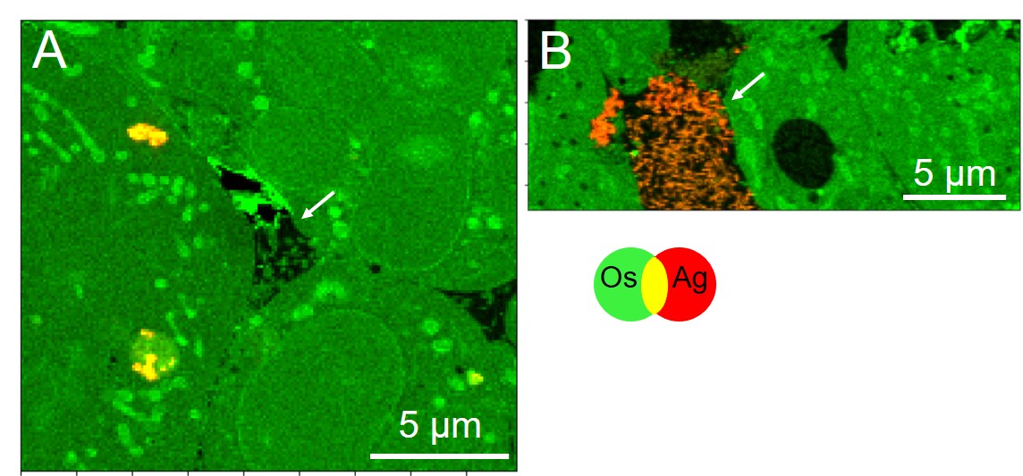
Figure: Nano-XRF imaging data of hepatocyte spheroids exposed for 7 days to citrate-coated silver nanoparticles (A) or to AgNO3 (B).