The
microelectronics industry is facing a physical limitation, and
will need to increase the density of
integrated components. One solution
could be to integrate microelectronics in three dimensions, as current
microelectronic circuits are planar. Stacking the components on top of each
other is one way to continue making them denser. This raises a new challenge: to
connect the components together once they are stacked.
Biologists and physicists from the CEA-iRTSV, the CEA-Leti, the CNRS, the UJF
and Inra at Grenoble decided to take advantage of the extraordinary
self-assembling capabilities of certain biological molecules, so that these
connections can build themselves. Many complex, regular structures in our cells
continuously assemble and disassemble. This is particularly the case for the
filamentous networks that make up the cell skeleton (cytoskeleton), composed of
actin.
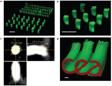
a)
3D visualization of two actin column networks of 1.5 (forefront) and
0.8 μm (behind), separated by 5 and 2 μm, respectively. b) 3D
visualization of a square array of actin micro-columns with a 0.5 μm
mesh. c) Horizontal and vertical slices of a micro-column showing
measurements averaged over one dozen structures. d) 3D visualization of a
network of polymerized actin from a microstructure representing the CEA
logo. Height of micro-columns: 36 ± 3 μm.
The researchers have developed a technique that enables controlling the
self-assembly of actin filaments in 3D, between 2 glass plates placed 30
microns apart and microstructured with a laser beam. The researchers then
injected a solution containing actin monomers between the two surfaces, which
polymerized in response to the geometry of the microstructures. As a result,
actin columns could be self-assembled in controlled shapes and sizes. Similarly,
the researchers have succeeded in making the columns grow from a surface into
hollow cylinders, produced on the other surface, much like male/female
electrical connectors. The connections were metallized with gold nanoparticles,
allowing an electrical current to pass between the two surfaces.
[1] Protein that comprises the skeleton of living cells and that can
regulate and control their form.