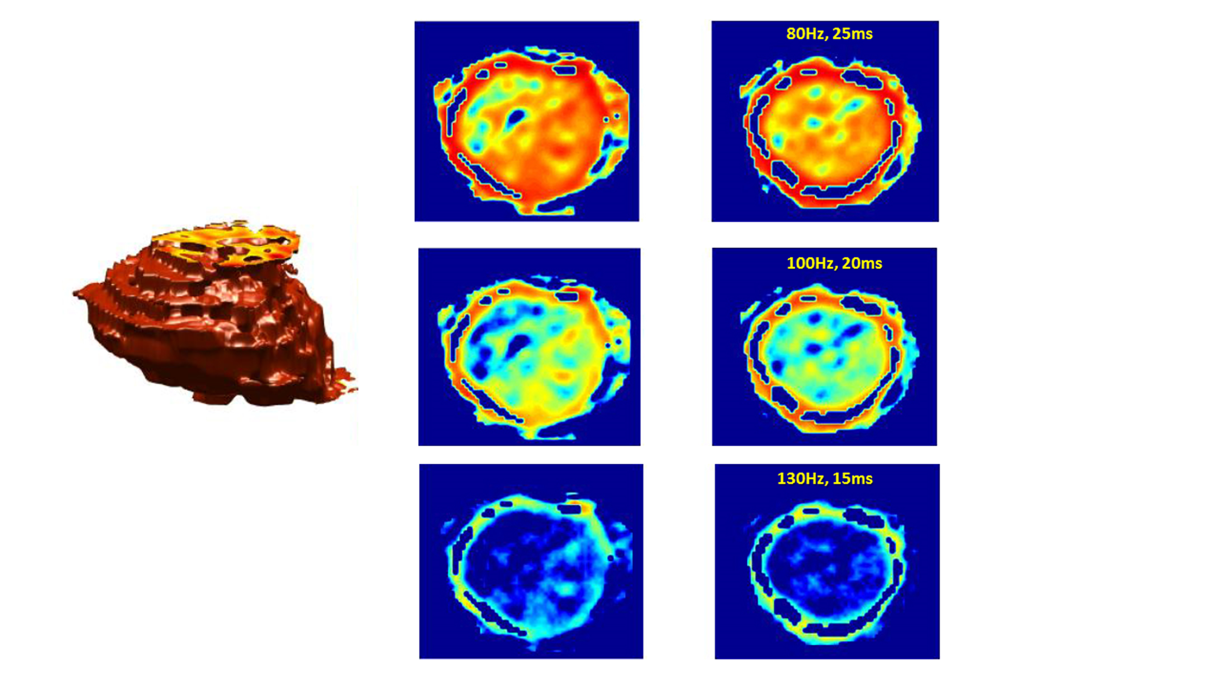
Conventional Magnetic Resonance Elastography (MRE) is used to identify lesions such as tumors or fibrosis, which are generally more rigid and less elastic than the surrounding healthy tissue. It is similar to a doctor palpating a patient... Yet much more sophisticated. Although very promising, this technique has a few issues: high exam cost, heavy installation for set up of each patient, low resolution of the images, instability of the elasticity estimation algorithm. Finally, it can only be used on superficial organs.
Noting that the elastic properties of tissues is closely linked to their microscopic structure (cell density, presence of fibers), the researchers tested the hypothesis that tissue elasticity can be directly estimated from images captured using diffusion MRI, i.e. based on the diffusion of water molecules rather than using mechanical vibrations. Developed in 1985 by Denis Le Bihan and his collaborators at the Frédéric-Joliot Life Sciences Institute, diffusion MRI (dMRI) is now widely used in medical imaging. This technique is very sensitive to tissue structure at the microscopic level.
The degree of liver fibrosis estimated through MRE and dMRI are 100% concomitant. This work has also pointed to a new type of contrast that can be produced from the elasticity images obtained by diffusion MRI. By simulating the passage of shear waves of any frequency or amplitude in the tissues, and without the technical difficulties of MRE, tissue structure heterogeneity can be revealed—in tumors in particular.
This result was the subject of a press release and a patent application by CEA.

Elastogramme virtuel 3D d'une tumeur du foie par IRM de diffusion et coupes à différentes fréquences de vibration (virtuelles) montrant l'évolution du contraste.
© CEA, Université de Yamanashi.