In recent years,
super-resolution microscopy (or nanoscopy), which uses optical microscopy to image biological samples at nanometric resolution, has undergone a real boom. Some nanoscopy methods use
photo-convertible fluorescent proteins* (PCFPs) as markers. One of the most widespread of these methods is Single Molecule Localization Microscopy (SMLM). It is based on the principle that PCFPs are able to change their fluorescence emission from green to red under illumination at 405 nm. But beyond this photo-conversion, PCFPs tend to switch reversibly between fluorescent and non-fluorescent states, which can seriously impact the quality of the images obtained. The teams of D. Bourgeois (IBS/I2SR) and B. Brutscher (IBS/RMN) have revealed a new photo-switching mode for mEos4b, a widely-used PCFP.
After photo-conversion, mEos4b (red form) undergoes a “negative photo-commutation”: it is switched off under light at 561 nm (which excites fluorescence) and reactivated under light at 405 nm. This property is due to a photo-induced conformational change in the chromophore (cis-trans isomerization), the cis conformation being the only one capable of fluorescence. The result is a marked blinking of single molecules during data collection.
Another, almost imperceptible, source of blinking was identified and elucidated in this study. Taking place over a shorter time scale (a few tens of ms versus several seconds for negative photo-commutation), a “positive photo-commutation” is observed: mEos4b in its red form is switched off under light at 405 nm and reactivated at 561 nm. The phenomenon can be explained using Nuclear Magnetic Resonance (NMR), which reveals conformational heterogeneity in mEos4b: a stable red fluorescent state (R1) predominates, but a second, much less bright red fluorescent state may appear (R2).
Light at 405 nm induces the transition from the R1 to the R2 state, while light at 561 nm restores the R1 conformation. The study also revealed that the coexistence of the two states R1 and R2 is strongly influenced by the physico-chemical conditions of the sample (pH, buffer medium, etc.).

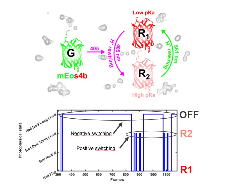
Top: mEos4b's positive photo-switching mechanism. Bottom, example of the evolution of the photo-physical state of a single mEos4b, molecule over time, showing transitions between the R1, R2 and OFF states. (D. Bourgeois / IBS)
Thanks to numerous developments, SMLM microscopy is now a key technique for integrative biology, making it possible to go from the cell to the single molecule.
Although numerous methodological obstacles have been overcome to improve the resolution of the images obtained, the study reveals that the great complexity of the photo-physical properties of fluorescent proteins is a limitation to the quantitative analysis of these images. However, there are still a number of solutions open to optimize experimental conditions and overcome this new lock.
Extra Information
photo-convertible fluorescent proteins*: fluorescent proteins whose fluorescence properties change irreversibly under illumination conditions.
Collaboration
- Irig/SyMMES/Équipe Conception d’Architectures Moléculaires et Processus Electroniques (CAMPE)
Fundings
- iNEXT-Discovery (871037) funded by the Horizon 2020 program of the European Commission,
Agence Nationale de la Recherche (grants no. ANR-20-CE11-0013-01, ANR-22-CE13-0046 and ANR-22-CE11-0011-01),
IR INFRANALYTICS (FR2054) FRISBI (ANR-10-INBS-0005-02) and GRAL, CBH-EUR-GS (ANR- 17-EURE-0003)