Measles virus is a highly contagious human pathogen that is currently on the rise in many countries, including France. The RNA genome of this virus is packaged (encapsulated) in helical molecular suprastructures, the nucleocapsids, consisting of thousands of nucleoproteins that bind to the entire genome of the virus. To spread in a cell, this virus uses a viral RNA polymerase. How does this polymerase access genetic material when it is protected by a capsid?
This is the task that IRIG researchers have been working on. In a previous study
[1], they had developed experimental methods to
in vitro encapsulate a given RNA sequence They had thus demonstrated that they could trigger self-organization of measles virus nucleoproteins into nucleocapsids i
n vitro by addition of RNA. They thus provided a simple and versatile tool for studying the mechanisms and rates of assembly of nucleocapsids, and allowed them to detect a clear dependence on RNA sequence.
In this new study
[2], they determine by cryo electron microscopy the three-dimensional high-resolution structure (3.3 Å) of these nucleocapsids. These structures reveal the positions and nature of the fine interactions between RNA and nucleoprotein. The identification of amino acids that are directly involved in encapsulation provides a better understanding of the stability of the superstructures, and their overall structure shows how the viral RNA polymerase is able to replicate the viral genome.
These new data could eventually lead to the development of new therapeutic strategies.
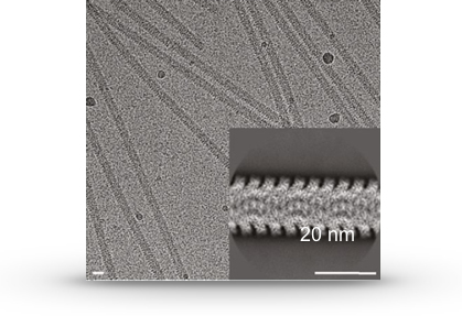 High resolution image obtained by cryo electron microscopy of nucleocapsids.
An average of these structures obtained by image analysis is also shown at the bottom left.
High resolution image obtained by cryo electron microscopy of nucleocapsids.
An average of these structures obtained by image analysis is also shown at the bottom left.