You are here : Home > Scientific news > New strategy reveals the organization of proteins within photosynthetic membranes

Highlight | Scientific result

New strategy reveals the organization of proteins within photosynthetic membranes


​​​​​​​​​​​​​​​​How do thylakoid membrane proteins organize themselves and work together to enable efficient photosynthesis? Researchers at CEA-Irig, from the BGE and LPCV laboratories, have successfully mapped these interactions in their native state. This breakthrough opens up new avenues for the integrative study of functional membrane systems, far beyond photosynthesis.

Published on 3 September 2026

​Photosynthesis is a process essential to life on Earth: it enables photosynthetic organisms (plants, algae) to convert light energy into chemical energy that can be used by the cell. Its efficiency depends in particular on the three-dimensional organization of numerous proteins present in the thylakoid membranes*. To understand how these proteins assemble and function together, researchers are developing proteomics approaches* to identify proteins that interact with one another. Among these approaches, cross-linking* coupled with mass spectrometry (XL-MS)* makes it possible to detect protein-protein interactions and reconstruct their networks. In this study, researchers at CEA-Irig developed an innovative strategy to map the protein networks of functional photosynthetic membranes.

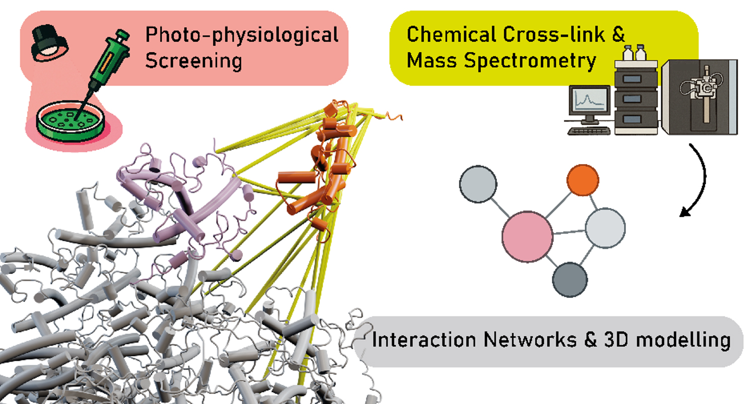
The researchers optimized the method based on cross-linking coupled with mass spectrometry (XL-MS) by combining the cross-linking agent, PhoX*, with a chemical agent, TMPAC* (trimethylphenylammonium chloride), to obtain reliable results while preserving the functional integrity of the membranes. This strategy enabled the identification of several hundred interactions—increasing their detection rate by 20 to 40 percent—between proteins involved in the regulation of photosynthesis. By combining these data with artificial intelligence (AI) modeling, the researchers revealed previously unknown associations involved in the tuning and repair of the photosynthetic machinery, thereby providing a precise understanding of the organization of the molecular complexes responsible for the conversion of light energy.​


© CEA-Irig/LPCV/P. Albanese
Figure : Improved chemical cross-linking approach to decipher protein networks in thylakoids membrane.

This research demonstrates that the method developed bridges the gap between functional and structural biology in plants by enabling the systematic exploration of protein networks under conditions close to physiological conditions. This approach provides a methodological framework that can be applied to other membrane systems—which are often difficult to study—paving the way for the modeling of new functional protein complexes.

Thylakoid membranes*: membrane structures located inside chloroplasts, organelles in plant cells (and algae) responsible for photosynthesis. The initial stages of photosynthesis (capturing light and converting it into chemical energy) take place at these membranes.

Proteomic approaches*: analytical techniques used to identify and quantify proteins in cells, biological fluids or any other sample containing proteins. 

Cross-linking* (XL): a technique based on the use of a chemical reagent capable of creating a covalent bond between two proteins that are sufficiently close to each other. This approach allows their interactions to be frozen so that they can be investigated for a better understanding gained of how these proteins organize and function together. 

Mass spectrometry (MS)*: an analytical technique used to identify and quantify proteins. When combined with cross-linking (XL-MS stands for Cross-Linking Mass Spectrometry), it enables the detection of chemically linked proteins and the reconstruction of the protein network’s organization within a biological system. 

PhoX*: a negatively charged cross-linking reagent, which may limit its access to membrane surfaces that are also negatively charged, such as those of thylakoids. The addition of TMPAC*, a positively charged compound, reduces these electrostatic repulsions and improves PhoX’s accessibility to membrane proteins, while preserving the structural integrity and biological activity of the membranes.

UMR : Irig/PCV-LPM (UGA-CNRS-CEA-INRAe) ; Irig/BGE-EDyP (UGA-CEA-Inserm).

Fundings : ANR (PHOTO_DYN), Union Européenne (PhotoLINK, Chloro-Mito, PlanktON), ProFI.

Collaborations : UGA, CNRS, CEA, INRAe, Inserm.​


Top page