Some of human cells are endowed on their surface with motile threadlike appendices. This is the case in our bronchi, where the beating movement of cilia drives the flow of mucus, a protective barrier naturally present in our body. Other cells, like spermatozoa, show a single flagellum. The flagellum, whose structure is shared with that of the cilium, can also deform to create an undulatory movement, responsible for the “swimming” of spermatozoa.
FOCUS: the flagellum, a system combining filaments with molecular motors
A flagellum is made up of a structure of parallel filaments, known as microtubules. Molecular motors - dyneins - form links between the filaments. By actively putting themselves under tension, they force the filaments to slide along one another. When well-coordinated, this movement creates regular and rhythmic undulations of the flagellum. Researchers have sought for decades to understand how the filaments and molecular motors manage to organize themselves to create this movement that’s regular in time and space.
An innovative study system
ased on the work of the CytomorphoLab team at the CEA in Grenoble, it has become possible to polymerize, or “construct”, actin filaments
3] and organize them into networks of prescribed architecture.
The “Active mechanosensitivity of hair cells in the inner ear” team led by Pascal Martin, CNRS research director at Institut Curie, was using this actin assembly method when a remarkable phenomenon was observed “Mathieu Richard, then a PhD student in my team working on a different subject, tried to assemble actin filaments in a bath containing myosin motors, in order to test their activity. Surprisingly, the filaments moved close to one another and spontaneously formed
undulating flagella-like bundles," explains the researcher. It is the first time that we have observed this phenomenon in an “artificial” actin and myosin system.
In this system, the researchers use neither the microtubules nor the dynein of the flagella, but another type of filament - actin - and another molecular motor, namely myosin. However, the fact that they observe an undulatory movement very similar to that of a flagellum suggests that
common physical laws may apply to these different filament-motor systems.
Another surprising observation helped clarifying the undulation mechanism. Marie Pochitaloff, during her PhD in Pascal Martin’s team, visualized the distribution of the myosin along the filament bundles. Pascal Martin explains: “We expected to find most of the myosin at the base of the bundles since it is there that the number of filaments, and thus the number of potential connection sites, are the most numerous. Against all expectations, we found instead that the myosin attached where there were fewer filaments but where the curvature of the structure was the largest, before moving toward the tip of the bundle.”
This coupling between the curvature wave and the location of the motors reveals a close relationship between the curvature of the filament bundles and their binding to the molecular motors.
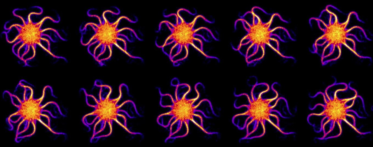
Tracking of the undulation of actin bundles over a period of time.
© Pascal Martin/Institut Curie
Between experimental biology and theoretical physics
Thanks to Martin Miranda’s work in theoretical physics, during his PhD supervised jointly by
Prof. Frank Jülicher at the Max Planck Institute for the Physics of Complex Systems and by Jean-François Joanny, Professor at Collège de France, it has been possible to
quantitatively model the experimental observations to understand the physical origin of the spontaneous undulation. “It is this collaboration between experiment and theory, between biology and physics, that made the results possible”, adds Pascal Martin.
This approach opens a new avenue to study, both experimentally and theoretically, the complex phenomena involved in the self-organization of filament-motor systems. It has already revealed a correlation between the curvature of filament bundles and their binding to molecular motors, as well as the existence of general physical laws in these system. Future studies will aim at clarifying the molecular mechanisms that allow the motors to feel the curvature of the filaments, and how it is possible to regulate this mechano-sensitivity.
This work constitutes a first step towards better understanding of the function and malfunction of cilia and flagella, and beyond that, of other biological processes involving deformations of the cytoskeleton by molecular motors.
[1] The work was conducted in
-
the Physical Chemistry Curie Lab (UMR168 - Institut Curie, CNRS, Sorbonne University) by the “Active mechanosensitivity by hair cells in the inner ear” team led by Pascal Martin;
-
at the Max Planck Institute for the Physics of Complex Systems (Dresden, Germany) by the team of Prof. Franck Jülicher, in collaboration with Prof. Jean-François Joanny (Institut Curie/Collège de France);
-
at the Cell & Plant Physiology Laboratory in the CytomorphoLab team, headed jointly by Laurent Blanchoin and Manuel Théry (CNRS, CEA, UGA, INRAE).
[2] The artificial flagellum created here is the result of the assembly of a large number (several thousand) of individual molecules.
[3] The actin filaments, like the microtubules, are one of the essential components of the cytoskeleton, allowing the cell to adopt various shapes and move under the action of molecular motors.