Elastography, in echography, is a method for measuring the elasticity (also known as hardness) of biological tissues. It is based on the very precise measurement of the propagation rate of shear waves induced in tissues by focusing an ultrasound beam. The speed of these waves is directly related to the elasticity of the tissue in which they propagate. Measuring this elasticity, or shear modulus, is increasingly used to diagnose various pathologies, such as breast cancer (a breast lesion is often harder than the surrounding healthy tissue) or liver fibrosis (during which the liver becomes increasingly hard). In this way, elastography makes it possible to avoid a large number of liver biopsies, or to reclassify breast cancer diagnoses. However, some cancers are not well characterized by elastography, and new biomarkers need to be identified to complement these measurements.
In this study, the authors focused on the nonlinear shear modulus, which refers to the fact that a soft medium, such as biological tissue, changes hardness under the effect of an external constraint. By compressing the organ under study with the ultrasound probe during an elastography measurement, it is possible to monitor this new biomarker and potentially refine the doctor's diagnosis. The aim of the present work was to validate such a measure of nonlinearity in a collaborative study using echography, magnetic resonance imaging (MRI) and a numerical simulation method. More specifically, the aim was to validate results obtained with in vitro echography on a phantom (a material simulating biological tissue) using two other approaches, one experimental, MRI elastography, and the second numerical, finite-difference simulation. For MRI elastography, they developed an innovative method that enabled them to measure the nonlinear modulus by imposing an external constraint on the phantom placed inside the imager. The results were confirmed by the numerical approach developed in collaboration with a team from INSA Lyon, which takes detailed account of the theory of acoustoelasticity (taking into account variations in the stiffness of a soft medium under uniaxial constraint). All three methods led to a similar measurement of the nonlinear modulus.
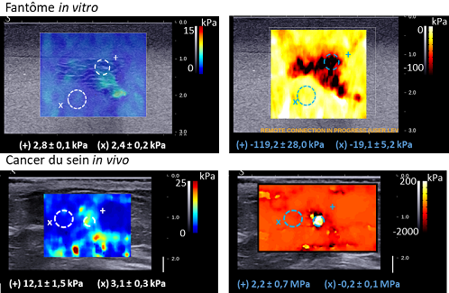
Measurement of elasticity (shear modulus, left) and measurement of nonlinear elasticity (nonlinear shear modulus, right) on an in vitro phantom (piece of liver in an agar-gelatin block, top) and in the in vivo breast in the presence of a cancerous tumor (bottom). The contrast of the nonlinear modulus is much better in both cases and allows good determination of tissue contours for diagnosis (from Bernal et al., In Vivo Quantification of the Nonlinear Shear Modulus in Breast Lesions: Feasibility Study, IEEE-UFFC, 2015)
This work validates the measurement of nonlinearity by clinical imaging methods (echography and MRI) and paves the way for its use as a new biomarker with particular interest in the diagnosis of breast cancer pathologies.
Contact : Jean-Luc Gennisson (jean-luc.gennisson@universite-paris-saclay.fr)
Ultrafast ultrasound elastography is a technique that records the propagation of shear waves generated by focused ultrasound. Tissue elasticity is directly related to wave propagation speed.