Team Leaders
Vincent Dive
vincent.dive@cea.fr
Radiolabeling
CEA has a long-term tradition in radioactivity manipulation that is maintained and developed in SIMOPRO for the labelling of peptides, pseudopeptides and proteins with tritium, but also with 14C and recently with 99mTc.
This activity gives rise to multiple collaborations including both with academic and industrial partners.
In vivo imaging.
Radioimaging
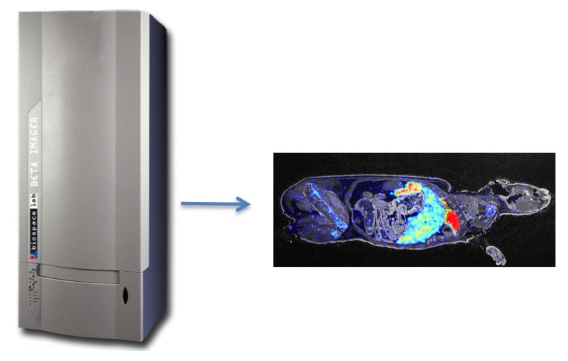
Labelled of biomolecules with stable isotopes such 3H and 14C are useful to establish in preliminary experiments either pharmacokinetics and tissue biodistribution of potential therapeutic molecule. The latter objective can be assessed by using digital radioimagers, like those developed by the Biospace company, the beta-imager.
The beta-imager allows real-time imaging, with extreme counting sensitivity (0.007 cmp/mm2 for 3H and 0.01 cmp/mm2 for 14C) and absolute signal quantification.
Optical Imaging
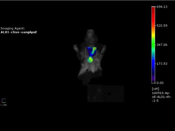
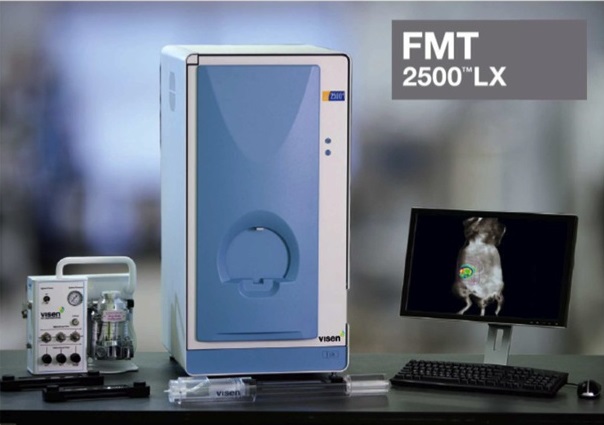
Peptides and proteins are labelled with near-infrared fluorescent group allowing their detection in vivo by 3D-fluorescent imaging system (FMT-2500 from Perkin Elmer).
Animal facility (mouse, rat & rabbit)
Performed in a dedicated animal facility, the most common experiments in SIMOPRO are:
- Microsurgery,
- minipumps implantation for perfusion of ligands in therapeutic applications
- development of mouse tumor models (syngenic, xenograft and spontaneous) and atherosclerosis models
- frozen tissue sections for radioimaging studies.
- intratracheal installation and aspiration methods for lung exposure studies.