Imagerie de la plaque instable (Contrat NIH, collaboration avec l’université de Yale, Dr M. Sadeghi).
Imaging of unstable plaque (NIH Contract collaboration with Yale University, Dr. Sadeghi).
From RXP470.1 compound, various imaging agents can be developed (Fig. 9).
Issuance of anti-inflammatory agents with functionalized nanoparticles by RXP470.1 (Athero-Nano Project LABEX LERMIT).
Nanoparticles that are surface decorated with RXP470.1 compound can be developed to target in the atherome plaque the MMP-12 present at the macrophages surface and thereby allow the local delivery of anti-inflammatory drugs (Fig. 10).
Injected into ApoE -/- mice, such nanoparticles can be detected by fluorescence within atheromatous plaques (Fig. 11, collaboration with our LABEX LERMIT colleagues (Prof.E. Fattal, Dr S. Lesieur and Dr. Doris E.).
The RXP470.1 as a new antiviral agent : MMP-12 plays a key role in the response to viral
- Allowing particularly IFN-alpha secretion
- inactivating INF-a
Thus, treatment of mice infected with a virus via RXP470.1, preventing INF-a inactivation, leads to a more rapid clearance of virus, indicating that the RXP470.1 compound represents another generic antiviral agent.
New generation of MMPs inhibitors
The RXP470.1 structure simplification leads to very potent compounds, even in absence of phosphoryl group (compound 14, Fig. 13).
A new mode of interaction
The compound 19 crystallographic structure (Fig. 14) shows that the carboxylate group in the side chain of the glutamate residue is interacting with the zinc atom, behaving as the chelating group in this compound.
MMPs active forms Detection
Synthesis inhibitors having photoactivatable chemical groups and radioactive atoms allows forming covalent complexes after irradiation facilitating detection of these complexes (Fig. 15).
Principle of detection from serum albumin functionalized with potent MMPs inhibitors.
Modified "serum albumin" protein is used to capture the MMPs active forms, which can be identified with a secondary antibody (Figure 16).
Halogenated operating in the development of MMPs selective ligands
The use of halogen (F, Cl, Br and I) to control the selectivity of MMPs substrates, in particular vis-à-vis the MMP-9 (Fig. 17).
X-bond interaction
The crystal structure analysis allows to detect the formation of an interaction between iodine and a carbonyl group of the protein involving a water molecule, X-bond interaction type (Fig. 18).
Study of the carbon nanotubes translocation, 14Carbone labeled
After pulmonary delivery of nanotube to mice can be observed after several months translocation of 14C nanotubes in various organs through the use of the radioimaging (Fig. 19).
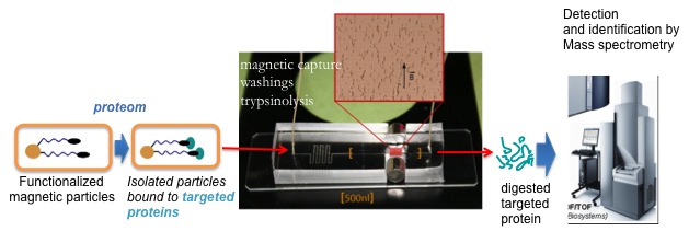
Project microfluidics in partnership with the CEA Material Sciences Directorate
In the context of CEA's TECSAN and of the DigiDiag program Investments for the Future program, the laboratory has developed in close collaboration with the group of F. Malloggi (DSM/UMR 3299 CEA/CNRS NIMBUS-LIONS), a microfluidic chip for proteomics studies.