The
exchange of ions across neuronal membranes gives rise to an
“electric discharge”, namely an action potential that determines the passage of
nerve impulses from one neuron to the next. The issue for microscopic imaging is
to follow the dynamics of these exchanges along neuronal circuits in different
biochemical conditions. Such studies are opening a new field of investigation to
cognitive neuroscience and drug development. Thanks to NeuroSpin’s
high-field
MRI device (17.2 Tesla) dedicated to small animals, teams from the
CEA-I
2BM are developing imaging techniques in living cells to bridge
the results between biological imaging on tissue sections and
in vivo
imaging. The first offers a spatial resolution of about 10 microns [1], while
the second has a resolution from 100 – 200 microns.
The researchers worked with Aplysia, a classic model animal in
neuroscience with a small set of large neurons (from several hundred microns to
1 mm in size). The neurons are therefore also observable by high-resolution
in vivo imaging. The scientists have shown for the first time that the
dynamics of transporting manganese ions (analogous to calcium) in the
motor neurons of the Aplysia buccal ganglion are modulated by the
neurotransmitter dopamine. To do this, they used high-field MRI to observe
Aplysia neurons in medium with a low manganese concentration, so as to
not disrupt cell physiology. They showed that stimulation of neurons with
dopamine alters the dynamics of manganese ions, particularly their elimination
by neurons. This is the first time that such a modulation is revealed in a
network where each neuron is visible by MRI, thanks to its very advanced spatial
resolution.
The exchange of ions across neuronal membranes gives rise to an
“electric discharge”, namely an action potential that determines the passage of
nerve impulses from one neuron to the next. The issue for microscopic imaging is
to follow the dynamics of these exchanges along neuronal circuits in different
biochemical conditions. Such studies are opening a new field of investigation to
cognitive neuroscience and drug development. Thanks to NeuroSpin’s high-field
MRI device (17.2 Tesla) dedicated to small animals, teams from the
CEA-I2BM are developing imaging techniques in living cells to bridge
the results between biological imaging on tissue sections and in vivo
imaging. The first offers a spatial resolution of about 10 microns [1], while
the second has a resolution from 100 – 200 microns.
The researchers worked with Aplysia, a classic model animal in
neuroscience with a small set of large neurons (from several hundred microns to
1 mm in size). The neurons are therefore also observable by high-resolution
in vivo imaging. The scientists have shown for the first time that the
dynamics of transporting manganese ions (analogous to calcium) in the
motor neurons of the Aplysia buccal ganglion are modulated by the
neurotransmitter dopamine. To do this, they used high-field MRI to observe
Aplysia neurons in medium with a low manganese concentration, so as to
not disrupt cell physiology. They showed that stimulation of neurons with
dopamine alters the dynamics of manganese ions, particularly their elimination
by neurons. This is the first time that such a modulation is revealed in a
network where each neuron is visible by MRI, thanks to its very advanced spatial
resolution.
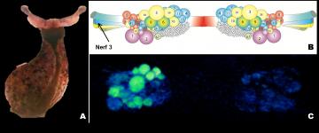
A.
Aplysia californica. B. Schéma du ganglion buccal. Le code couleur
relie les corps cellulaires aux nerfs périphériques qui reçoivent leurs
axones. C. Rendu 3D d’images par résonance magnétique, après migration
du manganèse le long du nerf 3. Les neurones qui ont accumulé du
manganèse (en vert) sont ceux avec des projections axonales dans le nerf
3
[1] 1 micron= 10-6 mete