Magnetic resonance imaging (MRI) is a non-invasive technique used for the acquisition of anatomical images and functional data. « Conventional » MRI is based on the visualization of protons of the water contained in tissues. However, when the contrast between healthy and pathological tissues is low, it is difficult to obtain usable data. In order to improve image quality, contrast agents such as gadolinium are often administered which, although effective, have limitations (lack of specificity, not directly quantifiable, potential toxicity).
In order to obtain a specific and directly quantifiable MRI signal, contrast agents containing, in their structure, atoms directly detectable in MRI, such as fluorine (there is practically no endogenous fluorine in living organisms), are used. Several probes for 19F MRI exist, such as PERFECTA (suPERFluorinatEd ContrasT Agent), unfortunately very poorly soluble in water, which limits its in vivo applications.
This is in this context that the SCBM Nanosciences team has developed, in collaboration with a team from UMR BAOBAB (NeuroSpin), micellar nanometric vectors for diagnostic purposes for the targeting of tumors and their visualization by MRI of fluorine 19 in vivo. These amphiphilic vectors incorporate a surface chemistry that allows their passive accumulation in the tumor area and a central reservoir for the encapsulation of a perfluorinated probe (PERFECTA), easily detectable by MRI. The development of an adapted MRI sequence has thus made it possible to visually delimit, with very good sensitivity, the volume of the tumor mass in model mice and to precisely quantify the accumulation of micellar nano-vectors in different organs (Figure).
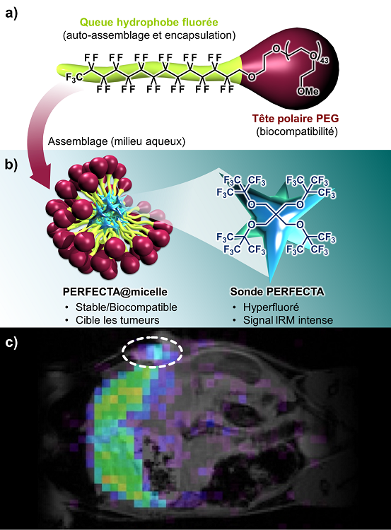
These results demonstrate the possibility of monitoring in real time the accumulation of micelles in tumor tissues according to an Enhanced Permeability and Retention (EPR) mechanism. This is the first example of the use of such a nano-vector encapsulating the PERFECTA probe.
Contacts : edmond.gravel@cea.fr; sebastien.meriaux@cea.fr;
eric.doris@cea.fr