In vivo 1H or 31P MRS allow for the non-invasive detection and quantification of more than 20 biomolecules (or metabolites) involved in energy metabolism (phosphocreatine, ATP, lactate, glucose), neurotransmission (glutamate, GABA), neuronal (N-acetyl-aspartate) or glial (myo-inositol, glutamine) function/density or membrane metabolism (choline compounds, phosphoethanolamine). Thus, a metabolic profiling of the brain can be performed longitudinally to explore and follow metabolic alterations in pathological conditions.
Following the administration of 13C-labeled energy substrates such as [13C]Glucose, [13C]Acetate or [13C]Lactate, detection of carbon-13 nuclei in biomolecules using dynamic 13C MRS allow for the direct assessment of rates of metabolic pathways. By following the incorporation of the 13C nuclei from these energy substrates through the intermediates of the tricarboxylic acid cycle (TCA), it is possible to estimate the rates of the neuronal (VTCAn) and glial TCA (VTCAg) cycles as well as the recycling rate of glutamate corresponding more or less to the rate of glutamatergic neurotransmission (VNT). This is done by adjusting the 13C fractional enrichments time-courses via the numerical resolution of the differential equations derived from a metabolic model of the neuronal-glial functional unit. No other method offers the opportunity to investigate specifically the neuronal and glial compartments as well as the glutamatergic neurotransmission.
Figure 2. Dynamic 13C NMR Spectroscopy.
Sodium-23
yields the second strongest NMR signal among biologically relevant NMR-active
nuclei. Yet, the average concentration of Na+ ions in the brain
remains 1/2000 that of protons. Furthermore, only the low intracellular 23Na
concentration (ISC~15 mmol/L) is considered as an operational proof of cellular
integrity and viability. As a consequence, 23Na MRI has been
proposed as a relevant imaging approach for the early detection of brain
disorders such as brain tumours, ischemic stroke, Alzheimer’s disease and
multiple sclerosis. A sophisticated method of multiple-quanta-filtering (MFQ)
takes advantage of the differences in mobility of the Na+ ions in
the intra- and extra-cellular compartments to filter out the signal from the
extracellular space. By combining the MFQ preparation module to a fast imaging
encoding schemes, maps of ISC can be obtained. The implementation of such tool
at 11.7T could complement advantageously our other cerebral imaging methods to
probe cellular metabolism and homeostasis.
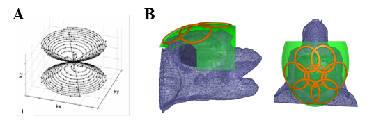 | Figure 3. 3D Twisted-projection imaging acquisition scheme (A) and prototype of a 23Na RF coil dedicated to imaging the brain of non-human primate at 7T (B). TPI encoding scheme from Riemer et al. (MAGMA 2013). |
Finally, lithium salts are the leading treatment for relapse prevention in patients with bipolar disorders. However, its mechanism of action remains unknown. For years, psychiatrists have been looking for a non-invasive method to investigate the transport kinetics of lithium salts through the BBB and the subsequent spatial distribution of lithium in the brain, assuming that both aspects play a critical role in the understanding and the successful management of lithium treatment. Recently, Komorovsky et al. have demonstrated that 7Li MRSI could be used to study the brain of bipolar patients in a clinical setting. By harnessing the increased polarization at 7T, we are looking to improve the time and spatial resolution of such acquisitions in order to investigate in collaboration with Pr. Frank Bellivier (Inserm UMR-S1144, Centre Expert Bipolaire et Dépression Résistante de la fondation FondaMental, Hôpital Fernand Widal, Paris) the relationship between lithium brain distribution and its therapeutic response.