Huntington's disease (HD) is a rare, hereditary, neurodegenerative disease that appears during adulthood. Currently, there are no treatments able to slow or cure the disease.
The course of HD is marked first by a presymptomatic, clinically-silent phase followed thereafter by a symptomatic phase associating progressive motor, psychological and cognitive disorders.
The presymptomatic phase is poorly understood. Identifying early biomarkers would greatly contribute both to a better understanding of HD progression and the evaluation of the efficacy of future therapeutics.
Imaging studies can contribute to that goal by furnishing more-pertinent functional information in a noninvasive manner. Among the various technologies, because of its particularly good spatial resolution and the diversity of contrasts and parameters it is able to measure, magnetic resonance imaging (MRI) is particularly well-suited for biomarker research.
An earlier glutamate metabolic imaging (gluCEST) study done in aged HD mouse models revealed the increased vulnerability of the corpus callosum, a white matter structure connecting the two cerebral hemispheres.
Aiming to better understand the role of cerebral connections and white matter in HD, researchers from MIRCen's Neurodegenerative Diseases Laboratory partnered with colleagues from the University of Grenoble and University College London to perform a study on mouse models of HD. Specifically, The looked at HD model mice aged from 2 to 18 months so as to observe disease course in a time span corresponding to young adulthood to old age in humans.
For their study, published in Human Molecular Genetics, the team developed a multimodal¹ MRI protocol to identify not only anatomic modifications within the brain but also the metabolic and structural modifications appearing as the mice aged.
The results showed that defects appeared in the corpus callosum as early as five months of murine age with no accompanying clinical signs. Those defects also preceded metabolic modifications, which, according to the gluCEST imaging used to detect them, appeared at eight months of age in the corpus callosum and propagated thereafter to neighboring structures at twelve months. Of note, the team observed atrophy in the cortex and corpus striatum relatively belatedly, at 18 months of age. This is of interest because cortical and striatal atrophy is the main biomarker used to evaluate HD progression in humans. The vulnerability of the striatum and the frontal and motor cortices, when considered with the alterations to the corpus callosum, seems to suggest a potential role for white matter and the glutamatergic pathway in the cerebral dysfunction encountered in HD.
The clinical adaptation of the team's multimodal imaging protocol may enable the identification of more robust presymptomatic-phase biomarkers, empowering in turn improved evaluations of targeted therapies.
1 : associating structural imaging, glutamate metabolic imaging (gluCEST), diffusion tensor imaging and magnetization transfer imaging.
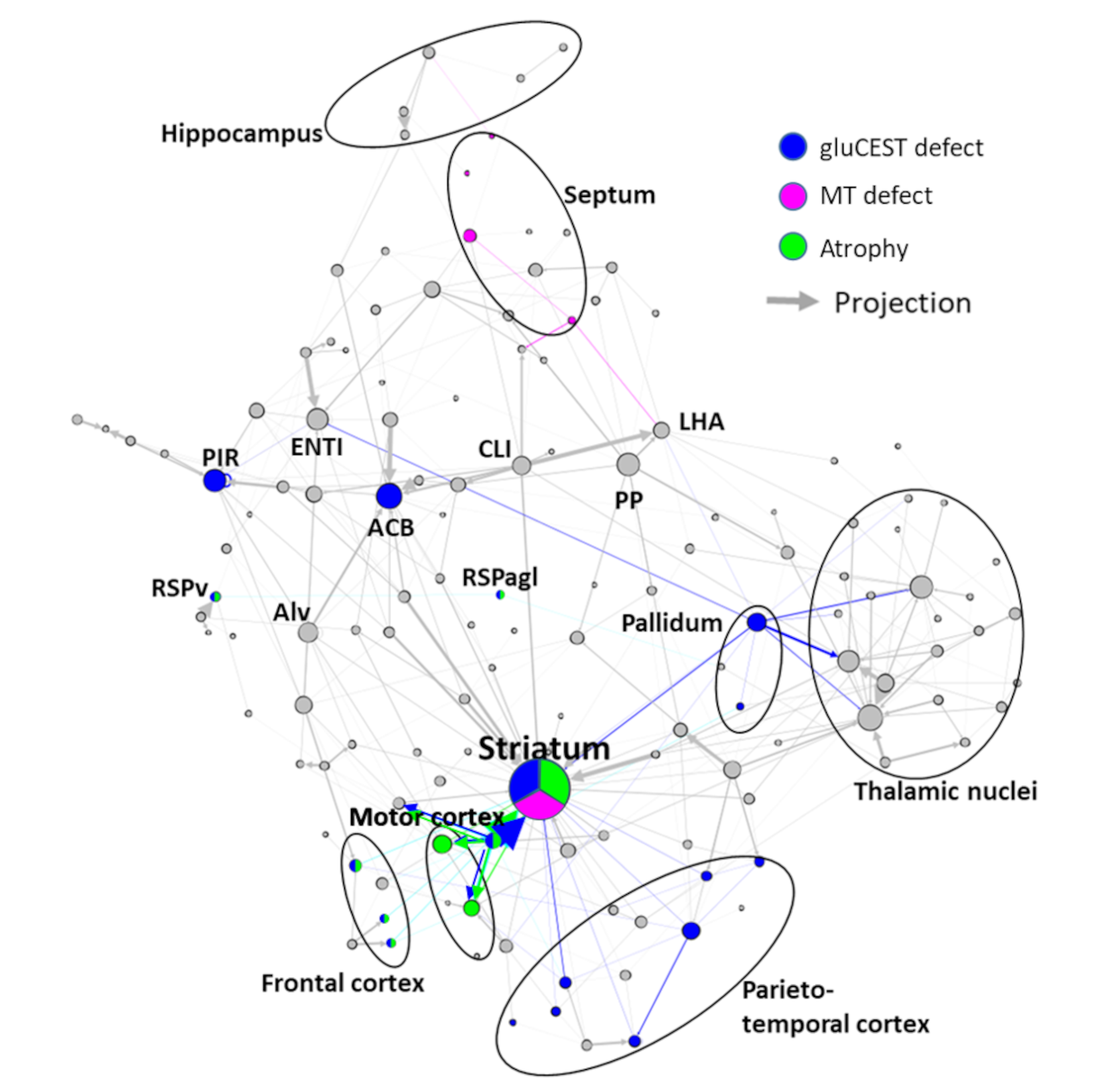
Figure :
Graph-theory representation of the main alterations to cerebral structures and their connections observed in the mouse model of Huntington's disease. The gray arrows and circles represent respectively axonal projections and regions. The colored circles indicate affected regions as determined in this study. The colored arrows indicate axonal projections between affected regions and may be associated with white matter alterations. (Credit: J. Flament)