Renewing scientific equipment: a challenge for French research
Neurodegenerative diseases suc as Parkinson’s, Alzheimer’s, Huntington’s, and multiple sclerosis, represent a major public health challenge and a priority field of investigation for translational research. In this context, the National Infrastructure in Biology and Health, NeurATRIS, is pursuing the renewal of its equipment to maintain cutting-edge research technologies. Since 2012, NeurATRIS has benefited from funding under the Programme Investissements d'Avenir to equip its core facilities with innovative technologies, particularly in imaging.
In 2024, NeurATRIS received additional funding through the France 2030 Investment Plan to replace its aging PET imaging systems. As a result, the MIRCen department acquired in July 2025 two new IRIS XL PET-CT cameras (Inviscan Imaging Systems). These systems replace the now-obsolete Focus 220 PET cameras and are the first operational IRIS XL units in France. The first images were obtained in October 2025.
Improved resolution and combined PET + CT imaging for a better understanding of neurodegenerative diseases in vivo
PET-CT technology combines two imaging modalities: metabolic imaging using positron emission tomography and anatomical imaging using computed tomography. It provides both anatomical images and information on the metabolic activity of cells.
The two IRIS XL cameras, equipped with a dual-ring PET detector system, are the first of their kind installed in France. This configuration provides an expanded axial field of view—from 104 to 400 mm—sub-millimeter resolution (< 1 mm), and improved sensitivity (≈ 4%). These new systems allow whole-body imaging in rodent models, as well as head and torso imaging in non-human primate models.
Whereas the Focus220 cameras lacked a CT system, the IRIS XL cameras generate CT images automatically aligned with PET images, sharing the same field of view, and used to compute attenuation correction for PET scans.
The system includes a fully self-shielded CT scanner, ensuring improved radiation protection for users and enabling rapid acquisition of high-resolution structural images (~70 µm) for PET attenuation correction, anatomical monitoring, or stereotactic guidance. The 3D reconstruction and segmentation tools ensure precise co-registration with MRI images acquired on the 7T (non-human primate models) and 11.7T (rodent models) core facilities at MIRCen.
The first images have been acquired in two models: rodent and non-human primate.
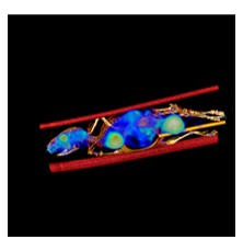 @N. Van camp / CEA
|
3D reconstruction of a whole-body CT acquisition superimposed on a 3D PET image. The CT scan shows the skeletal anatomy of the rodent, while the PET image (in color) displays the whole-body biodistribution of a radioligand. Organs in which the radioligand accumulates most intensely appear in colors tending toward red, while areas with low radioactivity are shown in dark blue. This study aimed to assess the biodistribution of a new PET radioligand across whole-body organs.
|
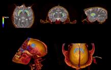 @ N. Van camp / CEA
| PET-CT images of an F18-labeled dopaminergic transporter and three-dimensional projection
|
These systems significantly strengthen MIRCen’s ability to study pathological mechanisms and the efficacy of innovative therapies for neurodegenerative diseases in vivo. They represent a strategic investment to accelerate the transfer of discoveries to the clinic for the benefit of patients.
Contact: Nadja van Camp