The limitations of current immunotherapies
Clear cell renal cell carcinoma (ccRCC) is the most common form of kidney cancer. These tumors are heavily infiltrated by immune cells, making them a particularly relevant target for immunotherapy. Antibodies blocking the PD-1/PD-L1 immune checkpoint have therefore profoundly changed the management of advanced forms of the disease.
However, their effectiveness remains limited: as the authors point out, only around 30% of patients show a tumor response to conventional immune checkpoint inhibitors. A better understanding of the diversity of immune cells found within tumors is therefore essential to identify new therapeutic targets.
The researchers focused on the HLA-G / ILT2 axis, another mechanism involved in immune regulation. HLA-G is a molecule that is normally absent from most adult tissues but is frequently expressed in a range of cancers, including kidney cancer. By binding to the inhibitory receptor ILT2 expressed by certain immune cells, HLA-G can restrain their activity and thereby contribute to tumor immune escape.
Two very different T-cell populations
Previous work by the team had identified two distinct populations of CD8⁺ T cells within renal tumors: cells expressing PD-1, which are well characterized and notably associated with a state of functional exhaustion; and a less well understood population expressing ILT2 but not PD-1.
In this new study, the researchers performed an in-depth characterization of these ILT2⁺ CD8⁺ T cells by combining several technologies, including high-dimensional spectral flow cytometry, single-cell transcriptomics, and T-cell receptor repertoire analysis. The study notably included tumor samples from 51 patients with clear cell renal cell carcinoma.
The analyses confirmed that ILT2⁺ cells represent a population that is markedly different from PD-1⁺ cells. They display a terminally differentiated phenotype and possess a powerful cytotoxic arsenal, including molecules required to kill target cells. Their profile within the tumor is also highly similar to that of their counterparts found in the blood, suggesting that they may be recruited from the peripheral circulation into the tumor.
“Bystander” T cells that are far from passive
One of the most striking findings concerns the antigen specificity of these cells. Unlike many tumor-infiltrating T cells, ILT2⁺ CD8⁺ T cells do not appear to be predominantly directed against tumor antigens.
Analysis of their T-cell receptors revealed, in particular, an enrichment of cells recognizing viral antigens. These cells therefore largely belong to the population of so-called “bystander” T cells: they are present within the tumor, but their TCR is not specifically directed against it.
At first glance, this observation might suggest that they play only a secondary role in the antitumor response. The study, however, shows the opposite.
The researchers demonstrate that these cells possess a killing capacity that can be activated independently of antigen recognition through their TCR. Following stimulation, they can use activating receptors more commonly associated with innate immune mechanisms to recognize and attack their target cells.
NKG2D: an alternative pathway for attacking tumor cells
Coculture experiments highlighted the role of the activating receptor NKG2D. This receptor enables ILT2⁺ CD8⁺ T cells to detect specific signals expressed by target cells and trigger their cytotoxic activity without relying on conventional TCR-mediated antigen recognition.
These “bystander” T cells therefore possess an alternative activation pathway that may confer antitumor activity. This makes them a particularly interesting population: highly cytotoxic cells that are already present within the tumor could potentially be mobilized against it, even when they do not directly recognize tumor antigens.
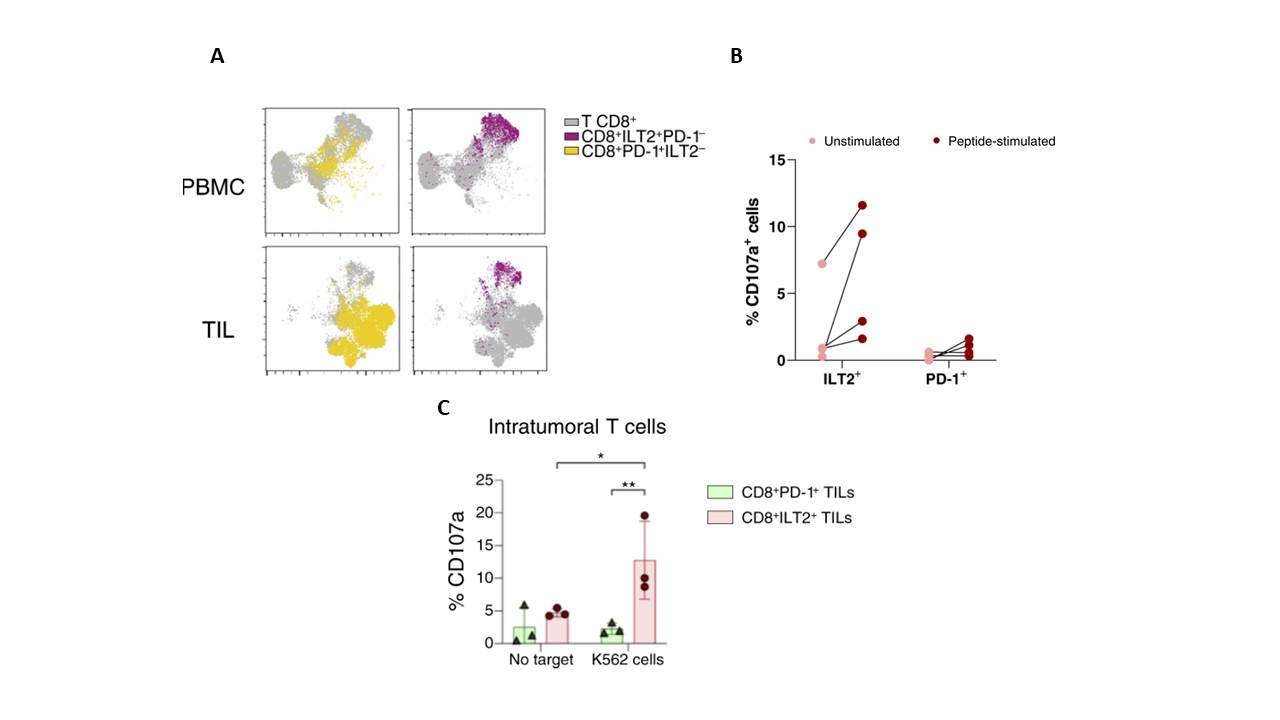
A. Spectral flow cytometry characterization of CD8⁺ILT2⁺PD-1⁻ and CD8⁺PD-1⁺ILT2⁻ T cells in peripheral blood and tumor samples from patients with ccRCC. B. “Bystander” CD8⁺ T cells infiltrate ccRCC tumors and express ILT2. ILT2⁺ CD8⁺ T cells display antiviral specificities identified by flow cytometry based on expression of the degranulation marker CD107a. C. Tumor-infiltrating CD8⁺ILT2⁺ T cells exert antitumor activity through a TCR-independent mechanism. © N. Rouas-Freiss / CEA
HLA-G restrains this cytotoxic potential
However, tumors also possess mechanisms that can neutralize this response. The researchers show that HLA-G expression by target cells inhibits the activity of ILT2⁺ CD8⁺ T cells.
The interaction between HLA-G and its receptor ILT2 therefore acts as a genuine immune checkpoint, capable of restraining a highly cytotoxic T-cell population. This observation further strengthens the therapeutic potential of targeting the HLA-G/ILT2 axis.
These findings provide a broader view of antitumor immunity. Rather than exclusively targeting T cells that specifically recognize cancer cells, it may also be possible to harness these “bystander” T cells present within the tumor microenvironment and endowed with potent cytotoxic activity.
Releasing the HLA-G/ILT2 brake, potentially in combination with the activation of mechanisms such as NKG2D, could therefore provide a basis for developing new immunotherapy strategies for patients with kidney cancer, particularly when treatments targeting PD-1/PD-L1 are insufficient.
Contact : Nathalie Rouas-Freiss