The "Imaging of Infection and Immunity" (L3i) Technological core laboratory of the IDMIT Department focuses on:
the dynamics of pathogen transmission and dissemination,
the dynamics of the host immune response to infection and/or treatment,
the distribution and impact of therapeutic interventions.
The laboratory aims to characterize the mechanisms of pathogenic microorganisms dissemination and the dynamics of immune responses, from the cellular scale to the whole-body level.
To this end, the laboratory is equipped with a variety of optical and nuclear imaging systems:
| - A clinical PET/CT system (Vereos, Philips®), combining positron emission tomography (PET) and computed tomography (CT), is available. This non-invasive in vivo imaging technique provides functional or metabolic information—after injection of radiotracers—as well as whole-body morphological data. This advanced technology is routinely used in hospital nuclear medicine and radiology departments, facilitating technology transfer from preclinical to clinical research. The installation of this system within a biosafety level 3 (BSL-3) laboratory enables its use for the investigation of a wide range of infectious and non-infectious diseases. Available radioisotopes: ¹⁸F, ⁶⁴Cu, ⁸⁹Zr, ⁶⁸Ga, ¹¹C
|
|
| |
|
|
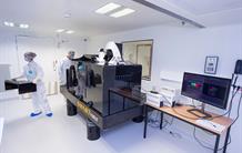
- A two-photon microscope specifically adapted for non-human primates imaging (Leica®, TCS SP8 MP) is also available. Installed within a biosafety level 3 (BSL-3) laboratory, this system enables in vivo 2D/3D visualization of pathogens, immune responses, and compounds of interest at the cellular scale. This is a unique equipment worldwide.
- A macroscopic fluorescence imaging system enabling in vivo detection and monitoring of locally administered fluorescent compounds. - A fibered confocal endomicroscopy device allowing in vivo visualization of fluorescent probes or pathogens, locally at the level of the skin or mucosal surfaces of the respiratory, gastrointestinal, or reproductive tracts.
| 
IDMIT two-photon microscope in BSL3 conditions
© L. Godart / CEA
IDMIT ex vivo confocal videomicroscopy system in BSL3 conditions
© L.Godart / CEA
|