A 0.5 Gy dose increases neural progenitor motility
During brain development, neural progenitor cells must proliferate, differentiate and migrate in a tightly coordinated manner to establish brain architecture. Disruption of their migration may therefore contribute to abnormalities in brain development.
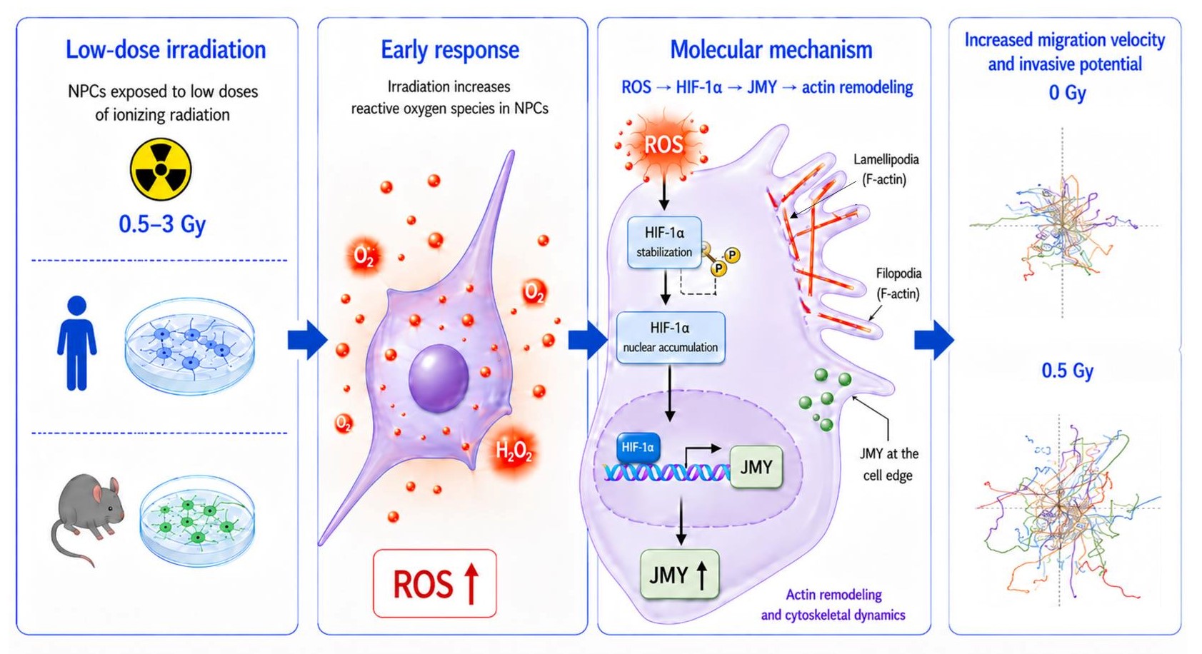
DRCM researchers investigated the effects of irradiation doses ranging from 0.5 to 3 Gy on human fetal and mouse neural progenitor cells. They show that a single exposure to 0.5 Gy is sufficient to rapidly increase cell migration speed. This effect, observed in vitro in several experimental models of migration and invasion, was also confirmed in vivo following intracerebral grafting of irradiated mouse cells, which showed increased dispersion into the surrounding brain tissue.
At this dose, the increase in migration was associated neither with premature differentiation nor with a specific phase of the cell cycle.
A ROS/HIF-1α/JMY cascade triggered by irradiation
The authors then sought to identify the molecular mechanisms underlying this response. Irradiation first induces an increase in reactive oxygen species (ROS). These promote the nuclear accumulation of HIF-1α, a transcription factor best known for its role in the cellular response to hypoxia.
HIF-1α then increases the expression of JMY, a protein capable of promoting actin nucleation and polymerization. Following irradiation, JMY is particularly enriched at lamellipodia and filopodia, actin-rich structures that are essential for cell movement.
Functional analyses confirm the role of this pathway: inhibiting ROS, HIF-1α or JMY prevents the irradiation-induced increase in migration and invasion. The authors therefore identify a ROS/HIF-1α/JMY signaling cascade that controls the migratory response of neural progenitor cells to irradiation.
Better understanding the effects of irradiation on the developing brain
These findings reveal an effect of irradiation that has been less extensively studied than cell death or altered proliferation: changes in the migratory behavior of neural progenitor cells.
Excessive or poorly controlled migration could disrupt cell positioning during brain formation and thereby contribute to some of the neurodevelopmental consequences associated with exposure to ionizing radiation.
The authors nevertheless emphasize that the pathophysiological consequences of this response will need to be further characterized in models that more closely reproduce brain development and allow its long-term effects to be investigated. A better understanding of this mechanism could also shed light on the processes involved in the recruitment of neural progenitor cells toward injured areas of the brain.
Contact : Laurent Gauthier